Researcher Mobility Support
Application code: RMS-24-01-04
Guest working organisation: Radiation and Nuclear Safety Authority of Finland (Säteilyturvakeskus – STUK)
Employing Organisation: Vinča Institute of Nuclear Sciences (VINS), National Institute of the Republic of Serbia, University of Belgrade
Research focused on the determination of the Half-Value Layer (HVL) in clinical mammography in laboratory and clinical setups.
Main quantity of interest in mammography is the mean glandular dose (MGD), calculated as the product of air kerma and three conversion coefficients. These coefficients depend on the breast tissue size, glandularity of the breast tissue and the beam spectra. Two of these coefficients depend heavily on the Half-Value layer (HVL) of the primary beam. Besides the air kerma measurements, this makes the HVL measurements essential in quality control.
International guidelines, protocols and IEC standards propose HVL measurements in narrow beam conditions (narrow field geometry), to minimize the influence of scattered radiation on the measurement. Quality control in mammography is typically performed by medical physicists or other experts in the clinics using ionization chambers (IC) or X-ray multimeters (XMM).
HVL is a quantity difficult to measure even in laboratory setup under controlled conditions. However, measurements in clinical setup could pose an additional issue given that the setup itself and measurement conditions have their limitations. These limitations could potentially lead to an HVL value different from its true value, which consequentially could lead to the wrong MGD estimation and, also introduce larger measurement uncertainties.
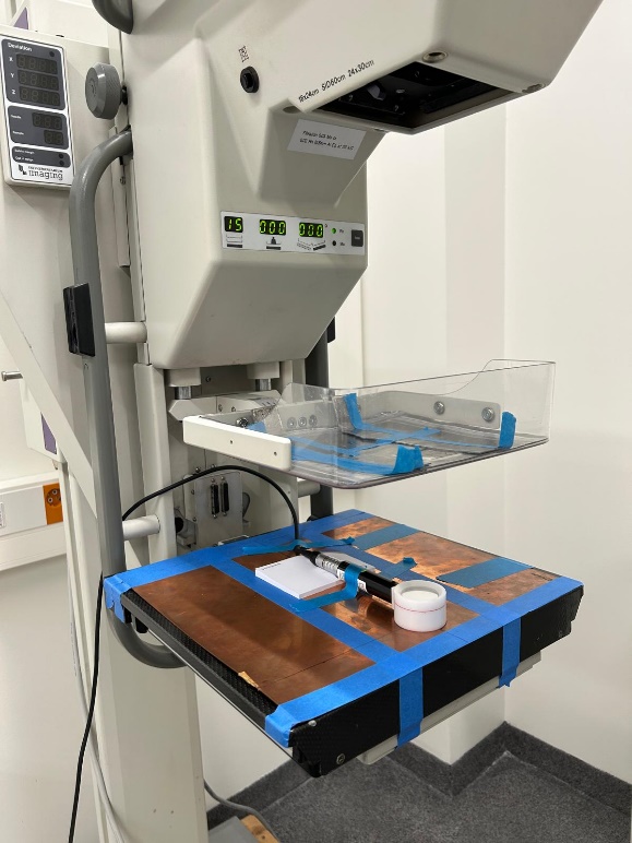
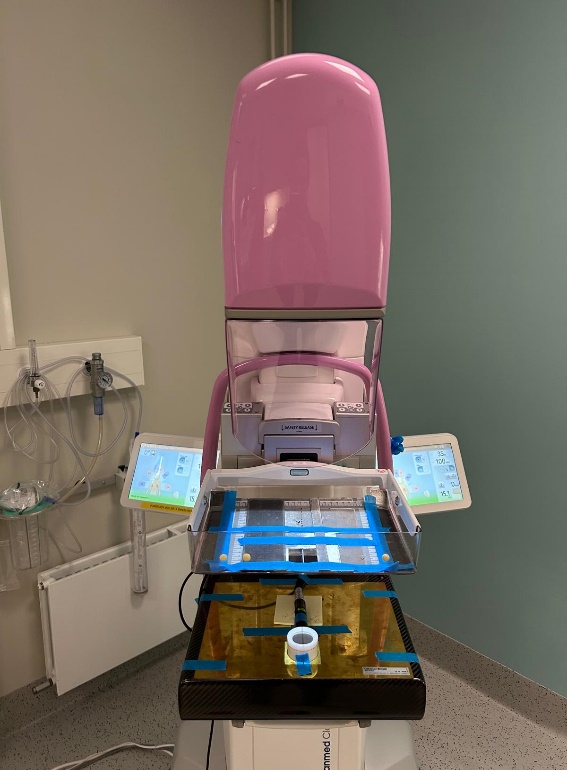
The study of this RMS focused on HVL measurements in mammography radiation fields different from the narrow beam conditions and, the impact of field size on HVL value and consequentially on estimation of MGD. Measurements were performed on two clinical mammography units: at Radiation and Nuclear Safety Authority of Finland (STUK) under laboratory conditions (Fig. 1) in Mo/Mo and Mo/Rh fields and at Helsinki University Hospital (HUS) under clinical conditions (Fig. 2) in W/Rh and W/Ag radiation fields.
Measurements were done with an IC and several XMMs in all radiation fields for tube voltages at 25 kV, 28 kV, 30 kV and 35 kV and for three different field sizes: open field without additional collimation of the primary beam and, two collimated fields of 10 cm x 10 cm and 5 cm x 5 cm size at the reference plane of the dosimeter. To mimic the realistic conditions, all measurements were done with the compression paddle in the primary beam on top of which the collimators were positioned. For radiation qualities in Mo/Mo and Mo/Rh fields, additional measurements were done without the compression paddle in the beam. The results were presented as the ratio of HVL values in open field and 5 cm x 5 cm field which is considered the smallest achievable realistic field in clinical setup.
Comparing the results, the impact of radiation quality was clearly observed. When the compression paddle is present in the beam, these relative differences in HVL values were higher compared to when the compression paddle was removed. This highlights the impact of forward scattering from the compression paddle on the results. When results are compared for the two anodes, Mo with lower HVL values and W with higher values, it is observed that the relative differences in measured HVL in open field and smaller fields are higher for the tungsten anode.
An extensive set of measurements was performed with an ionization chamber for additional field sizes for the reference radiation quality Mo/Mo at 28 kV defined in the existing guidelines and protocols. These additional fields were rectangular in shape and one had an irregular shape providing a 5 cm x 5 cm field at the reference plane of the IC but not collimating the whole beam, providing a better understanding of the geometry and field shape impact on measured HVL. In the case of rectangular fields, even when the total field areas were the same or similar, the shape of the collimated field plays a significant role. In case of the two fields that had the same collimation opening (5 cm x 5 cm covering the active area of the IC) but different coverage of the whole radiation beam, the influence of scattered radiation on the HVL result is seen and it is not insignificant. This information is crucial for clinical measurements, especially when smaller collimators not covering the whole area of the compression paddle (or the radiation beam) are used.
For several radiation qualities, spectrometry results, which are considered the zero-field measurements, were already available at STUK laboratory and were compared to the results in other field sizes, leading to even higher differences in measured HVL.
The XMMs are less sensitive to radiation scattered from the back and the sides, and for this reason they are not expected to be much dependent on the field size therefore, the measurements were done only in open field and the 5 cm x 5 cm field. The results show that the differences in these two field sizes were insignificant but when the results are compared to the HVL values measured with the IC these differences are higher. Even though the field size impact is smaller for XMMs compared to the IC, the larger difference between the XMM and the IC values for the same field size is highlighting the importance of correct XMM calibration in clinically relevant radiation qualities and, appropriate selection of radiation quality in the XMM software settings.
Key Achievements:
The results of this RMS lead to a conclusion that the HVL value measured with an IC can be significantly influenced by the field size, shape (including the geometry and dimensions) and the radiation quality, up to 5 % or even higher if compared to the spectrometry results. This directly impacts the estimation of MGD up to 5 % or even more. It is concluded that the partial collimation of the radiation beam also has a significant impact. It is seen that field size has a bigger impact for higher HVL values and, the differences in measured HVL values are more pronounced in the case of W anode.
When the field dimension is varied across the direction parallel to the chest wall edge, the difference in HVL values becomes more prominent compared to the dimension varied across the direction parallel to the X-ray tube.
Publications and Future Standards:
According to the results, harmonized measurements procedures and methods should be proposed to improve the accuracy of HVL measurements in mammography.
Based on the results of this RMS, a paper titled „Influence of radiation field size on HVL measurements in mammography“ was submitted to a peer-reviewed journal.
Dissemination of the work done was also done by two conference papers titled „Determination of the Half-Value Layer (HVL) in mammography: impact of field size” and “Determination of HVL in mammography using different methods“.